Lorem ipsum dolor sit amet, consectetur adipiscing elit. Integer vel eros volutpat, consequat diam ac, eleifend dolor. Mauris risus ante, tempus in interdum elementum, consectetur id odio. Praesent lorem dolor, sollicitudin sed metus at, laoreet vestibulum dolor.
CARSWELL, Robert (1793–1857)
Pathological Anatomy. Illustrations of the Elementary Forms of Disease.
London, printed for the author, and published by Longman, Orme, Brown, Green, and Longman, 1838 [1833–1838].
-
More info

-
More info

-
More info

-
More info

-
More info

-
More info

-
More info

-
More info

Related items
-

DAGONET, Henri (1823–1902)
Nouveaux traité élémentaire et pratique des maladies mentales suivi de considérations pratiques sur l’adminstration des asiles d’alienés.
Paris, J. B. Baillière & Fils, 1876. -

DU BOIS-REYMOND, Emil (1818–1896)
Untersuchungen über thierische Elektricität. Vol. I-II:1-2.
Berlin, G. Reimer, 1848-1860 (1884). -

AUVERT, Alexandre (d. 1865)
Selecta praxis medico-chirurgicæ quam Mosquæ exercet. Typis et figuris expressa Parisiis moderante Ambr. Tardieu. Pars I-III (of IV).
Parisiis, apud J.-B. Baillière / Hectorem ... -

CARSWELL, Robert (1793–1857)
Pathological Anatomy. Illustrations of the Elementary Forms of Disease.
London, printed for the author, and published by Longman, Orme, Brown, Green, and Longman, 1838 [1833–1...
First edition of this monumental pathological atlas, regarded as one of the finest ever produced. “The illustrations have, for artistic merit and for fidelity, never been surpassed, while the matter represents the highest point in which the science of morbid anatomy had reached before the introduction of the microscope” (DSB). Robert Carswell of Paisley, Scotland, was during his medical studies in Glasgow distinguished for his skill in drawing and was employed by Dr. John Thompson of Edinburgh to make a collection of drawings illustrating morbid anatomy. In order to fulfill this task Carswell spent two years (1822-23) at the hospitals of Paris and Lyon. After taking his M.D. at Aberdeen in 1826 he returned to Paris and resumed his studies in morbid anatomy under the celebrated P. C. A. Louis. In 1828 he became professor of pathology at the University College in London, but before assuming his teaching duties he was commissioned to prepare a collection of pathological drawings. He accordingly remained in Paris until 1831, during which time he had completed a wonderful series of two thousand watercolour drawings of diseased structures. – ”The great difficulty, and frequently the impossibility of comprehending even the best descriptions of the physical or anatomical characters of diseases, without the aid of coloured delineations, induced me to undertake the publication of the present work.” (Carswell) Back in London Carswell occupied himself with the preparation of his great book on pathological anatomy, the plates for which were furnished from his large store of drawings. From the 2000 watercolour images he had made, Carswell chose 48 to illustrate his pathological atlas. Each of these he drew himself on the lithographic stone and also coloured each plate by hand. The beautiful plates were published with accompanying text in twelve fascicles, beginning in January 1833. In view of the time-consuming work required in colouring the figures on each plate by hand, the edition is supposed to have been limited to 300 copies, which would make a total of 14 400 plates during a period of five years!
Collation: Pp (218), with 48 hand-coloured lithographed plates.
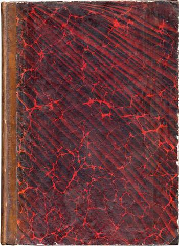
Binding: Swedish-bound half calf, gilt decorated spine, lemon yellow endpapers.
References: Garrison-Morton 2291; Long, History of Pathology (1928), 154-55; Heirs of Hippocrates1501; Notable Medical Books from the Lilly Library, 189; Osler 2250; Haskell, Norman Library 408; Hagelin, Rare and Important Medical Books, KIB, 166-67. Christie’s Norman Sale 968; Jeremy Norman Cat. 27 (1993), No. 56; Jeremy Norman Cat. 25 (1993), No. 87.