Lorem ipsum dolor sit amet, consectetur adipiscing elit. Integer vel eros volutpat, consequat diam ac, eleifend dolor. Mauris risus ante, tempus in interdum elementum, consectetur id odio. Praesent lorem dolor, sollicitudin sed metus at, laoreet vestibulum dolor.
VICQ D’AZYR, Felix (1748–1794)
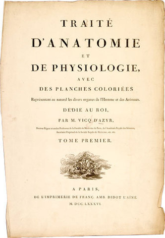
Traité d’anatomie et de physiologie, avec des planches coloriées représentant au naturel les divers organes de l’homme et des animaux. Tome Premiere [all published].
Paris, Franç. Amb. Didot, 1786.
-
More info
-
More info
-
More info
-
More info
-
More info
-
More info
-
More info
Related items
-
VICQ D’AZYR, Felix (1748–1794)
Traité d’anatomie et de physiologie, avec des planches coloriées représentant au naturel les divers organes de l’homme et des animaux. Tome Premiere [all published].
Paris, Fra... -
DARAN, Jacques (1701–1784) & GAUTIER D'AGOTY, Jacques Fabien (1716–1785)
Observations chirurgicales sur les maladies de l’urethre, traitées suivant une nouvelle methode. Nouvelle Edition.
Paris, chez Debure l’Ainé, 1748. -
NICHOLLS, Frank (1699–1778)
De anima medica prælectio ex Lumleii et Caldwaldi Instituto, in theatro Collegii Regalis Medicorum Londiniensium ad socios habita, die Decembris 16to. Anno 1748to. Editio Altera...
-
ALIBERT, Jean Louis (1768–1837)
Nosologie naturelle ou Les maladies du corps humain distribuées par familles. Tome Premier (all published).
Paris, Carpelet for Caille & Ravier, 1817.
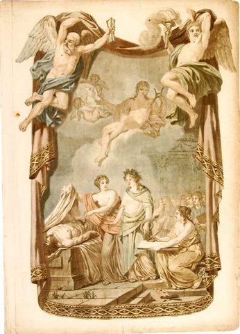
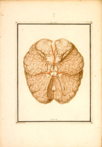
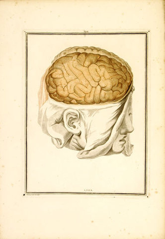
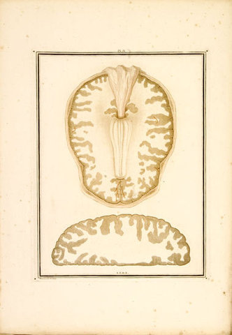
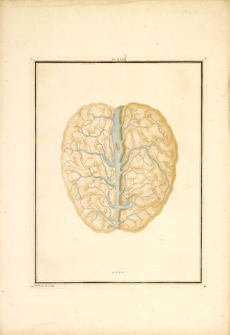
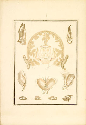
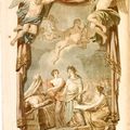
First edition of this outstanding anatomical folio atlas of the brain, the most accurate neuroanatomical work produced before the advent of microscopic staining techniques. The atlas has some of the finest coloured plates of the brain and nervous system that are to be found in neurological literature. The illustrations were at the time unrivalled in their quality as well as in their comprehensiveness. Complete copies are very rare. The work was issued in eight fascicles. The publication of the planned multi-volume work was interrupted by the French Revolution and the author’s premature death. The book was printed by François-Ambroise Didot, one of the most important members of the Didot family, who for several generations were the most prominent in French printing. The plates were drawn and engraved by Alexandre Briceau “dessinateur du Cabinet d’Anatomie” assisted by Mlle. Briceau. The work is dedidcated to Louis XVI, and the allegorical frontispiece shows the king as Apollo sitting on a cloud watching an anatomy scene with three goddesses around the corpse, one representing Study with an oil-lamp, one Medicine with a serpent, and the third the draughts-woman. Vicq d'Azur, permanent secretary to the Paris Academy of Medicine and personal physician to Marie Antoinette, found that his dissections of the brain were facilitated by first hardening the brain in alcohol. He identified accurately for the first time many of the cerebral convolutions, along with various internal structures of the brain. He rediscovered the white line in the calarine cortex and described the mammillothalmic tract which still bears his name, as well as the central sulcus with the pre- and postcentral convolutions and insula twenty years before Reil and Rolando (McHenry/Garrison).
Collation: Pp. (6), 123, (1); Section I: pp (2), 17, (1); II: pp (2), 19-38; III: pp (2), 39-68; IV: pp (2), 69-87, (1); V: pp (2), 89-111, (1). Aquatint frontispiece printed in colour and finished by hand, one engraved leaf of explanation of the frontispiece, and 69 engraved plates numbered I-XXXV, consisting of 34 aquatint outline plates, some with stipple engraving, printed in two or more colours with hand-finishing, accompanied by 34 outline plates with figures serving as key to the explanation of the plates, and one outline plate [No. XVIII] copied from Samuel Thomas Soemmering’s De basi encephali (1778).
Binding: The plates and the text in their folded quires as issued have remained unbound inside a contemporary half calf case-binding with ties. [protective box 220503]
Provenance: Gustaf Retzius (1842-1919).
References: Garrison-Morton 401.2; McHenry/Garrison, 104-106; DSB, XIV, 14-17; Norman Library, 2150; Hagelin, Rare and Important Medical Books in the Library of the Karolinska Institute, 124-5; Christie’s Norman Sale II, 840, in modern binding. Jeremy Norman Cat. 29 (1995), No. 254. Waller 9953.