Lorem ipsum dolor sit amet, consectetur adipiscing elit. Integer vel eros volutpat, consequat diam ac, eleifend dolor. Mauris risus ante, tempus in interdum elementum, consectetur id odio. Praesent lorem dolor, sollicitudin sed metus at, laoreet vestibulum dolor.
BERRES, Joseph (1796–1844)
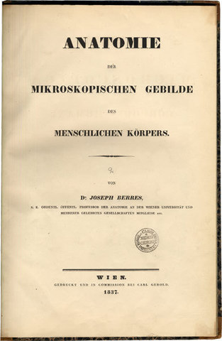
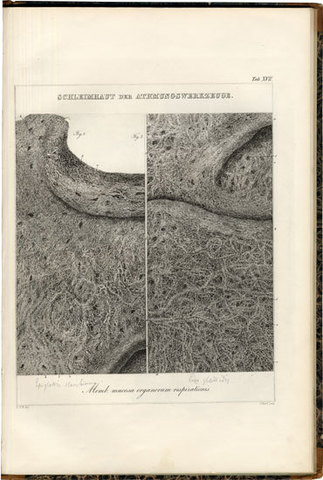
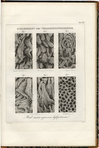
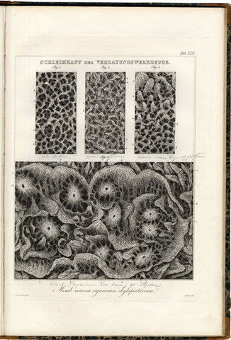
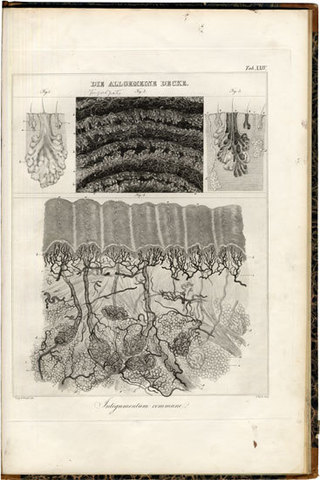
Anatomia microscopica corporis humani. – Anatomie der mikroskopischen Gebilde des menschlichen Körpers.
Viennae, typis Caroli Gerold, sumptibus auctoris, 1837 (1836-42).
-
More info
-
More info
-
More info
-
More info
-
More info
-
More info
-
More info
-
More info
Related items
-
DAGONET, Henri (1823–1902)
Nouveaux traité élémentaire et pratique des maladies mentales suivi de considérations pratiques sur l’adminstration des asiles d’alienés.
Paris, J. B. Baillière & Fils, 1876. -
SAYRE, Lewis Albert (1820–1900)
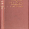
Spinal Diseases and Spinal Curvature. Their Treatment by Suspension and the Use of the Plaster of Paris Bandage.
London, Smith, Elder, & Co., 1877. -
MEYER, Édouard (1832–1902)
Traité des opérations qui se pratiquent sur l’œil.
Paris, H. Lauwereyns, 1871. -
VIRCHOW, Rudolf (1821–1902)
Die krankhaften Geschwülste. Dreissig Vorlesungen, gehalten während des Wintersemesters 1862–1863 an der Universität zu Berlin. Band I-III:1.
Berlin, August Hirschwald, 1863-[1...
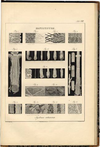
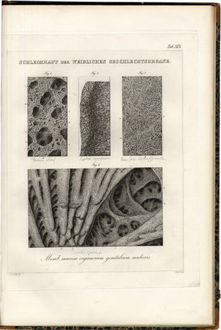
First edition of one of the earliest histological studies of the nervous system and one of the first to use microphotography, but the inscriptions on the plates do not indicate that any were made after photographs or daguerreotypes. Berres was the first to disclose a process of photographic etching, preceding Donné who kept his process a secret. Eder means that Berres probably was one of the first to use artificial light in microphotography, dating Berres’ first microphotograph to 1840, dating his disclosures to August, 1840. – ”As soon as Daguerre’s invention became known in Vienna, he [Berres] acquainted himself with this novel process and made use of it. . . . He obtained, as early as 1840, a microphotograph . . . he reproduced this by a process of etching daguerreotype plates, which he invented, and obtained prints with fatty inks on a printing press. He wrote about his experiments in the Vienna Zeitung of April 18, 1849.” (Eder) The beautiful plates are all drawn by Berres' students; Carl von Nagel, Christian Voigt, and Joseph Hyrtl, who all became great names in anatomy. Plates 2, 6, 7, 8, are lithographs, the others are engraved, most of them by Joseph Hyrtl. Berres was actually a surgeon, ”who after Mayer’s death obtained the Chair of Gross Macroscopic Anatomy at Vienna in 1830. His appointment to Vienna coincided with the time at which the Vienna optician Simon Plössl opened new possibilities of observation by his improved compound microscope. It was very tempting to try this out on Lieberkühn’s, Barth’s and Prochaska’s preparations in the anatomical museum. Berres thus became a microscopist. He began to study the tissues of the human body. In the new microscope he saw ”small bubbles” again and again and he reproduced them in his Anatomie der mikroskopischen Gebilde des menschlichen Körpers. This atlas developed into a truly wonderful work and the first in histology in general. Students of Berres, Josph Hyrtl, Carl Nagel and Christian Voigt, eagerly collaborated with him. Heliography could already be applied to duplicate the pictures and Berres himself invented a method whereby the photographs could be fixed by etching on a silver plate. Berres’ investigations had brought him close to one of the greatest discoveries which, however, was to be left to those who came after him (Schwann, Schleiden); Berres did perceive the cells and did reproduce them but he did not grasp what he saw. Still possessed by the theory of the fiber, hence still under the influence of Erasistratos (3rd century B.C.), Berres was able to see only ”cell bubbles” but could not recognize the cell in its particular structure.” (Lesky)
Collation: Pp (6) incl. title in Latin, 272; Atlas: pp (2) title in German, 24 (misprinted 21) with 24 plates.
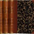
Binding: Two volumes, contemporary half calf with richly gilt spines and marbled boards.
References: Lesky, Erna, The Vienna Medical School of the 19th Century (1976), pp 73-4; Eder, History of Photography (1945), pp 386-87.