Lorem ipsum dolor sit amet, consectetur adipiscing elit. Integer vel eros volutpat, consequat diam ac, eleifend dolor. Mauris risus ante, tempus in interdum elementum, consectetur id odio. Praesent lorem dolor, sollicitudin sed metus at, laoreet vestibulum dolor.
BEALE, Lionel Smith (1828-1906)
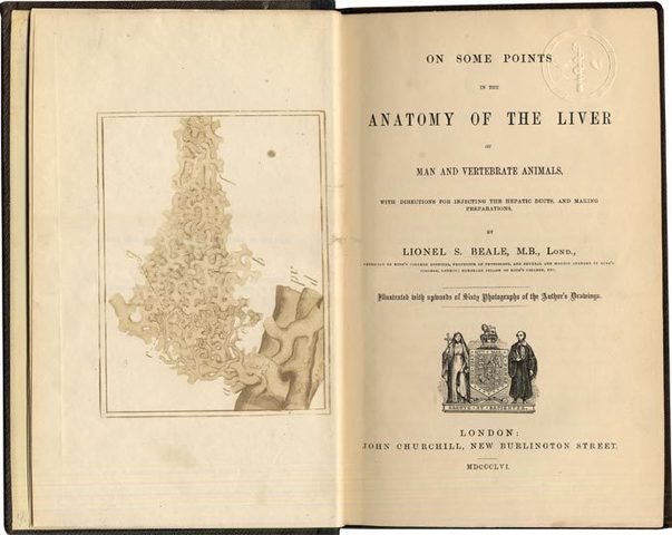
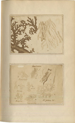
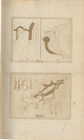
On Some Points in the Anatomy of the Liver of Man and Vertebrate Animals, with directions for injecting the hepatic ducts, and making preparations. Illustrated with upwards of Sixty Photographs of the Author’s Drawings.
London, John Churchill, 1856.
-
More info
-
More info
-
More info
-
More info
-
More info
-
More info
Related items
-
DAGONET, Henri (1823–1902)
Nouveaux traité élémentaire et pratique des maladies mentales suivi de considérations pratiques sur l’adminstration des asiles d’alienés.
Paris, J. B. Baillière & Fils, 1876. -
SAYRE, Lewis Albert (1820–1900)
Spinal Diseases and Spinal Curvature. Their Treatment by Suspension and the Use of the Plaster of Paris Bandage.
London, Smith, Elder, & Co., 1877. -
MEYER, Édouard (1832–1902)
Traité des opérations qui se pratiquent sur l’œil.
Paris, H. Lauwereyns, 1871. -
VIRCHOW, Rudolf (1821–1902)
Die krankhaften Geschwülste. Dreissig Vorlesungen, gehalten während des Wintersemesters 1862–1863 an der Universität zu Berlin. Band I-III:1.
Berlin, August Hirschwald, 1863-[1...
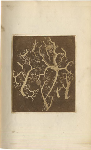
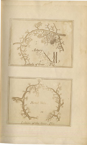
First edition. A rare book, of great interest as a very early example of a photographically illustrated medical book. Charles B. Wood knows of only one earlier example, the Quarterly Journal of Microscopical Science (1853) which contains one unmounted albumen print. The present work is much more substantially illustrated; it contains 14 leaves of photographs (albumen prints) with 26 drawings by the author, each containing smaller microscopical figures, giving a total of 66 numbered figures. Some of the negatives were made by Julius Pollock, but most were by Beale, who also made the prints in his labortatory at home. In the very interesting preface, Beale discusses the practicality of photographic book illustration, and gives data on the time and cost of producing prints. Of the edition size, Beale states that it was only “a few copies.” – “the photographs were taken from diagrams copied from the author’s drawings. Many of them have been much diminished . . . the diagrams from which the photographs were taken, were copied from drawings which had been traced from the preparations with the aid of the neutral tint glass reflector. The magnifying power has been estimated by comparison with the original objects, . . .” . Beale, one of the leading authorties on microphotography, was the first physiological investigator to practise the method of fixing tissues by injections. His discoveries included the pyriform nerve ganglion cells, called “Beale´s cells.” An unusually good draughtsman, Beale himself illustrated his books profusely with graphic drawings. His drawings of Beale’s cells are still reproduced in standard works on histology.
Collation: Pp. xx, 80, (32) adverts: Mr Churchill’s Publications. With 14 inserted leaves of albumen prints, one of which is placed as frontispiece.
Binding: Publisher’s cloth, outer hinges repaired, original spine with gilt lettering.
References: Not in Gernsheim’s Incunabula of British Photographic Literature (1984); DNB, Twentieth Century 1901-1911, vol. I, pp 118-120. Dobson, Anatomical Eponyms, p 26 “Beale’s Cells – nerve cells in the cardiac ganglia”. 14 Peter Eaton (booksellers) Ltd (sept. 2004).