Lorem ipsum dolor sit amet, consectetur adipiscing elit. Integer vel eros volutpat, consequat diam ac, eleifend dolor. Mauris risus ante, tempus in interdum elementum, consectetur id odio. Praesent lorem dolor, sollicitudin sed metus at, laoreet vestibulum dolor.
DUCHENNE DE BOULOGNE, Guillaume (1806–1875)
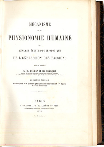
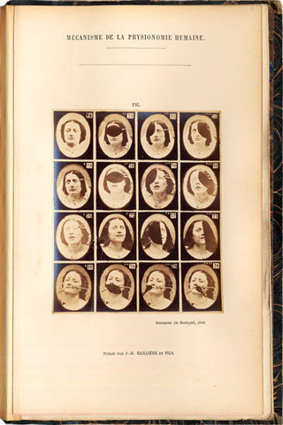
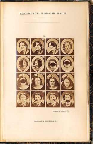
Mécanisme de la physionomie humaine ou Analyse électro-physiologique de l’expression des passions. Deuxième Édition accompagnée de 9 planches photographiées représentant 144 figures et d’un frontispice.
Paris, J.-B. Baillière et Fils, 1876.
-
More info

-
More info
-
More info
-
More info
-
More info
-
More info
-
More info
Related items
-
DAGONET, Henri (1823–1902)
Nouveaux traité élémentaire et pratique des maladies mentales suivi de considérations pratiques sur l’adminstration des asiles d’alienés.
Paris, J. B. Baillière & Fils, 1876. -
SAYRE, Lewis Albert (1820–1900)
Spinal Diseases and Spinal Curvature. Their Treatment by Suspension and the Use of the Plaster of Paris Bandage.
London, Smith, Elder, & Co., 1877. -
MEYER, Édouard (1832–1902)
Traité des opérations qui se pratiquent sur l’œil.
Paris, H. Lauwereyns, 1871. -
VIRCHOW, Rudolf (1821–1902)
Die krankhaften Geschwülste. Dreissig Vorlesungen, gehalten während des Wintersemesters 1862–1863 an der Universität zu Berlin. Band I-III:1.
Berlin, August Hirschwald, 1863-[1...
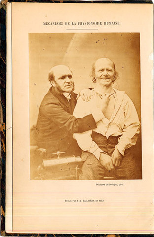
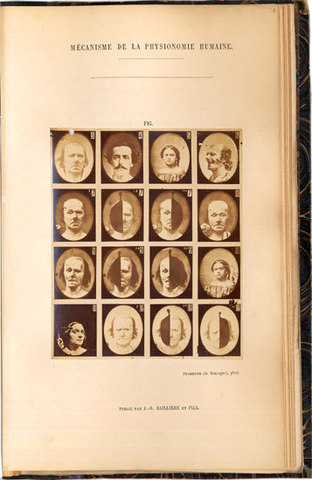
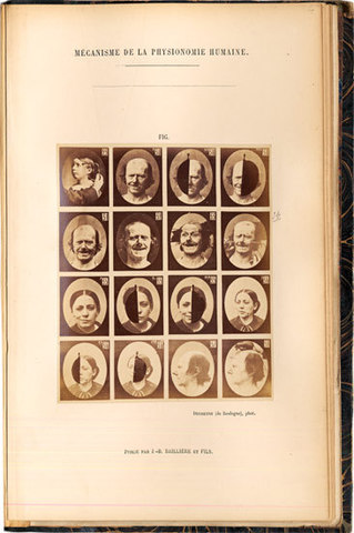
When first published in 1862 this pioneer work in physiological psychology was the first medical publication illustrated with photographs from life. The first edition was accompanied by an Album with ca 73 plates (the number of plates varies, no two are alike) each with a mounted photograph. This book was produced in a very limited number of copies and has been virtually unobtainable, copies remain only in a very few major libraries. This second edition of 1876 has the same plates as in the first: the famous frontispiece being a mounted albumen photograph with a self-portrait of Duchenne applying electrodes to the face of his patient to demonstrate the physiology of expression and 9 plates with mounted albumen photographs, each consisting of 16 reduced photos of patients’ faces, numbered 3-84 (figs. 1-2 are wood-engravings in the text). The work marks the beginning of photography applied to anatomy and neurology and Duchenne took all the photographs himself. Darwin later referred to Duchenne’s work and reproduced several of Duchenne’s photographs as woodcuts in his own Expressions of Emotions in Man and Animals (1872). In this famous work the great French neurologist brought together his understanding of facial anatomy and his skill in photography to describe and interpret the ways in which the face portrays the range and subtlety of human emotions. Using patients from hospitals in Paris as his subjects, he electrically stimulated their individual facial muscles and then photographed the results – the first time photography was employed to illustrate and record the results of a series of scientific experiments.
Collation: Pp xii, [v]-xi, (1) blank, 67, (1) blank, [1]-125, (1) blank, [129]-196. Three figures in the text (figs 1-2, 2 bis). Frontispiece and 9 plates with mounted photographs.
Binding: Contemporary dark green half morocco with five raised bands, marbled boards and marbled endpapers.
References: Garrison-Morton 4973 (1862 ed.); Grolier Club, The Truthful Lens 49; Gernsheim, Alison. ’Medical Photography in the Nineteenth Century’ in Medical and Biological Illustration, vol. IX, 1961. p 92; Jammes, André: ’Duchenne de Boulogne, La grimace provoquée et Nadar’ in Gaz. de Beaux Arts (1979); Cuthbertson, R.A. (ed.) The Mechanism of Human Facial Expression (1990); Sotheby’s, La Photographie II Collection Marie-Thérès et André Jammes (2002), Nos. 190-192; Gasser & Burns, Photographie et Médecine 1840-1880 (1991), Nos. 36-38; Thomas, Ann, Beauty of Another Order (1997), pp 134 ff; Waller 2607. Waller 2607.