Lorem ipsum dolor sit amet, consectetur adipiscing elit. Integer vel eros volutpat, consequat diam ac, eleifend dolor. Mauris risus ante, tempus in interdum elementum, consectetur id odio. Praesent lorem dolor, sollicitudin sed metus at, laoreet vestibulum dolor.
FOX, George Henry (1846–1937)
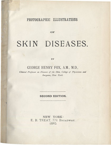
Photographic Illustrations of Skin Diseases. Second Edition.
New York, E.B. Treat, 1885.
-
More info
-
More info
-
More info
-
More info
-
More info
-
More info
-
More info

-
More info
-
More info
-
More info
-
More info
-
More info
Related items
-

DAGONET, Henri (1823–1902)
Nouveaux traité élémentaire et pratique des maladies mentales suivi de considérations pratiques sur l’adminstration des asiles d’alienés.
Paris, J. B. Baillière & Fils, 1876. -
SAYRE, Lewis Albert (1820–1900)
Spinal Diseases and Spinal Curvature. Their Treatment by Suspension and the Use of the Plaster of Paris Bandage.
London, Smith, Elder, & Co., 1877. -
MEYER, Édouard (1832–1902)
Traité des opérations qui se pratiquent sur l’œil.
Paris, H. Lauwereyns, 1871. -
VIRCHOW, Rudolf (1821–1902)
Die krankhaften Geschwülste. Dreissig Vorlesungen, gehalten während des Wintersemesters 1862–1863 an der Universität zu Berlin. Band I-III:1.
Berlin, August Hirschwald, 1863-[1...
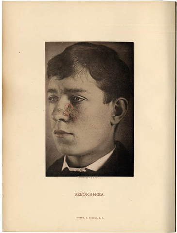
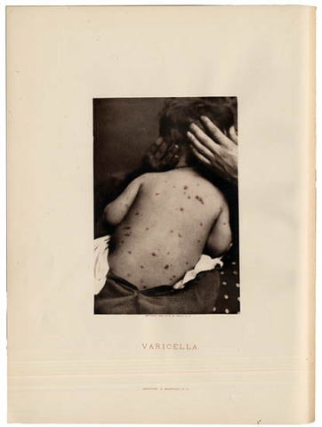
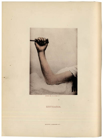
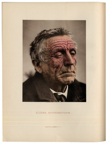
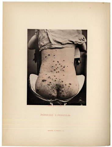
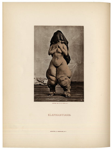
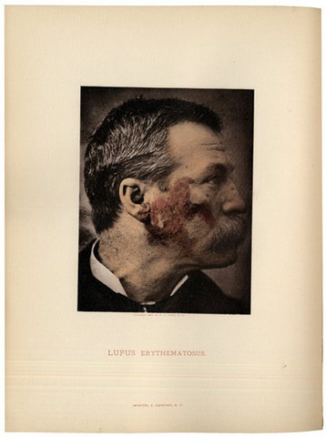
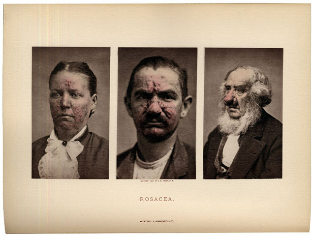
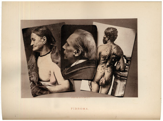
This is the second edition of “the first atlas in which the recent improved photographic processes have been employed in the portrayal of the diseases of the skin”. It was first published in 1880 with 48 plates. In this second edition “many imperfect plates have been replaced by new and better ones, and some single illustrations have been doubled or trebled in order to show different phases of a given disease. In the text, which has been increased to over two hundred quarto pages, special attention has been devoted to the diagnosis and treatment of skin diseases, and the work is now presented as a combined atlas and text-book. The plates are made from photographic negatives taken from life. The Artotype reproductions of these negatives are the work of Mr. Edward Bierstadt, and the hand-colouring of the plates has been entrusted to the well-known medical artist, Dr. Joseph Gærtner.” (Preface). Fox was professor at the Dermatological Clinic of the College of Physicians and Surgeons in New York. In 1902 together with Fordyce, he first described the Fox-Fordyce disease. Many of the photographs were taken by O.G. Mason at the Bellevue Hospital in New York City. The photographs show male and female patients with various skin diseases. “Acc. to Dann this was the first dermatological work to make use of a duplicating technique known as heliogravure or photo-engraving which was invented in 1878. This entails transferring a negative onto a copper plate by photomechanical means. The picture is thus etched onto the plate. The pictures all have the sharp focus of the original negative. Indeed they often surpass it in the richness of their shades of grey.” (Ehring)
Collation: Pp 208. With 48 plates with hand-coloured photographs reproduced in Artotype. Over 20 of the plates have more than one picture. 20th century blue cloth library binding.
Binding: Bound in are four chromolithographed plates not belonging to this work, showing Measles, Small Pox and Scarlet fever, from Morrow’s Atlas of Skin Disease, and one very fine lithograph plate taken from photo showing Tubercular Syphilide (Supplement to the Medical Record, Dec. 31, 1887, Lith. Lindner Eddy & Clauss, N.Y.)
Provenance: Library of the San Diego County Medical Society.
References: Garrison-Morton 3996; The Truthful Lens 60; Burns, Early Medical Photography in America (1983), p 1243; Heirs of Hippocrates 2094-95; Burns, A Morning’s Work (1998), plates 38, 46 and 48; Ehring, Skin Diseases, pp 223-24; Dann, Walther: Die Abbildung der Hautkrankheiten vom Beginn der modernen Dermatologie bis zum Ende des 19. Jahrhunderts. Inaug. Dissert. 1969.