Lorem ipsum dolor sit amet, consectetur adipiscing elit. Integer vel eros volutpat, consequat diam ac, eleifend dolor. Mauris risus ante, tempus in interdum elementum, consectetur id odio. Praesent lorem dolor, sollicitudin sed metus at, laoreet vestibulum dolor.
LYONET, Pierre (1707–1789)
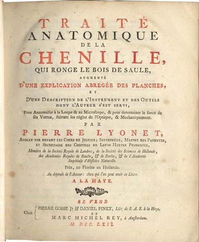
Traité anatomique de la chénille, qui ronge le bois de saule, augmenté d’une explication abregée des planches, et d’une description de l’instrument et des outiles dont l’auteur s’est servi, pour anatomiser à la loupe & au microscope, & pour determiner la force de ses verres, suivant les règles de l’optique, & mechaniquement.
A la Haye, chez Pierre Gosse, Jr. & Daniel Pinet et Marc Michel Rey, 1762.
-
More info
-
More info
-
More info
-
More info
-
More info
-
More info
-
More info
-
More info
-
More info
-
More info
-
More info
Related items
-
[FRANKLIN, Benjamin (1706–1790)]
Some Account of the Pennsylvania Hospital; From Its first Rise, to the Beginning of the Fifth Month, called May, 1754.
Philadelphia, B. Franklin & D. Hall, 1754. -
TREW, Christoph Jacob (1695–1769)
Abhandlung von einigen Verschiedenheiten welche an dem Menschen vor und nach seiner Geburt wahrgenommen werden, und den dabey sich äussernden Spuren der Allmacht und Weisheit Go...
-
EUSTACHIUS, Bartholomæus (c. 1510–1574)
Tabulæ anatomicæ ... Præfatione, Notisque illustravit, ac ipso suæ Bibliothecæ dedicationis die publici juris fecit Jo. Maria Lancisius.
Romæ, ex officina Typographica Francisc... -
SCHILLING, Gottfried Wilhelm (1725-1799)
De lepra commentationes. Recensuit J. D. Hahn.
Lugduni Batavorum, apud Sam. et Joan. Luchtmans, Trajecti ad Rhenum, apud Abr. van Paddenburg, 1778.
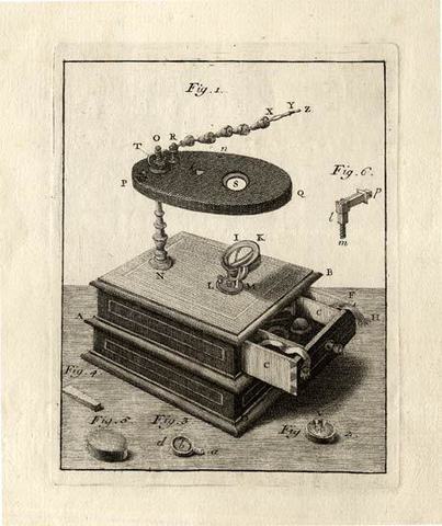
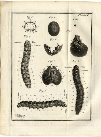
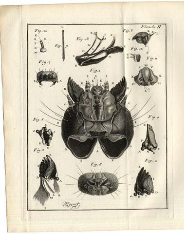
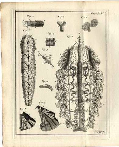
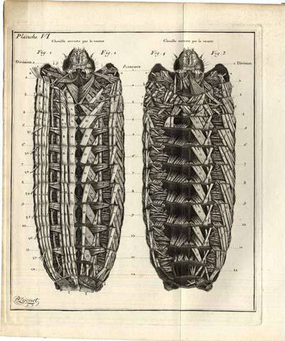
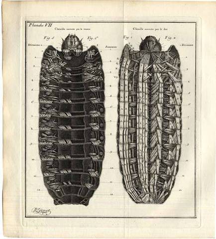
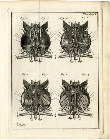
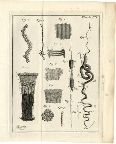
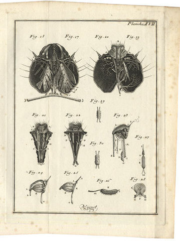
Second and the best preferred edition of this notable work with Lyonet’s beautiful preparations of the larva of the goat moth (Cossus ligniperda). The frontispiece is showing Lyonet’s microscope, which was used by Spallanzani. First published in 1760, this work is the fruit of immense labour and is rightly regarded as a classic in the field of comparative anatomy. Of the seventeen chapters, the most important are those which deal with the external and internal parts of the caterpillar, particularly the heart and muscles. Although the observations are minute and Lyonet’s drawings detailed and meticulous, Lyonet’s sensibilities prevented him from killing more than eight to nine of the creatures. “Lyonet examined the common goat moth caterpillar and the anatomy of its chrysalis and imago. He intended to delineate the metamorphosis of the insect in subsequent volumes, but was prevented from doing so by an affliction of the eyes, which after 1767 made it impossible for him to do close work. Traité anatomique is devoted wholly to the anatomy of the caterpillar, and the plates, drawn and engraved by Lyonet, portray the muscles, nerves, bronchia, heart, viscera, silk vessels, and the internal parts of the head with astonishing precision.” (DSB) – “Lyonet’s great monograph on the goat moth caterpillar remains today among the greatest examples of anatomical examination.” (Garrison-Morton).
Collation: Pp xxii, (2), 40 (explanation of the plates), 1-587, (3) index & avis au relieur, 589-616. With engraved frontispiece (Lyonet’s Microscope) and 18 folding engraved plates numbered I-XVIII, drawn and engraved by Lyonet and signed by him. Title printed in red and black.
Binding: Fine copy in contemporary red half sheep skin with sprinkled boards.
Provenance: 20th century label on front endpaper : “To Professor H. H. J. Nesbitt, Dean of the Faculty of Science, Carleton University. This token of friendly esteem, signed W. R. Thompson, Hon. D. Sc. Carleton University.”
References: Garrison-Morton 305; Cole Library 1714; Clay & Court, History of the Microscope, 59-60; Stuart Pierson in DSB, VIII, pp 579-80; Moe, Mikroskopets historie, pp 82-84. Waller 11895.